

By: Virgilio C. Ventura
Mental illness, though invisible, affects millions of people worldwide. Due to its intangible nature, it often goes unnoticed and misunderstood by society. Thus, the many ways of visualizing mental illness can aid in understanding its impact on individuals and society to break the stereotypes and stigma surrounding it.
Visual Artwork
One way to visualize mental illness is through artistic expressions. Artists have been utilizing their artwork as a medium for portraying mental illness in its unique forms. Contemporary artist Yayoi Kusama, who has lived with hallucinations and mental disorder all her life, depicts her struggles through her art. Her work ‘Infinity Mirror Room,’ through which a viewer is immersed in a seemingly endless field of multicolored lights, represents the obsessive and repetitive thoughts experienced by individuals with OCD. The surrealism of Salvador Dali’s work ‘The Persistence of Memory’ reflects the fragmented thoughts of people with schizophrenia.

The Cinema
Films have also been an effective medium to visualize mental illness. The movie ‘A Beautiful Mind’ depicts the story of John Nash, a renowned mathematician who lived with schizophrenia. The film vividly portrayed the unpredictable behavior and the visual and auditory hallucinations experienced by Nash. ‘Silver Linings Playbook’ tackles the struggle of a man with bipolar disorder, showcasing the highs and lows of the disorder with an honest and raw portrayal.

Information Graphics
Yet another way of visualization is through information graphics. Mental illness statistics, when visualized, can be a powerful tool to highlight the severity of the issue. Infographics can display facts like the growing number of people affected by depression, the demographic most affected, the sexes most susceptible, and the social, physical, and financial effects of the condition. The World Health Organization (WHO) periodically releases infographics on mental health issues to raise awareness.
Medical Imaging Technology
Likewise, one of the challenges in understanding and treating mental illness is its invisibility to the naked eyes. Specifically, in the case of dementia (a mental condition that affects the brains of 35.6 million people worldwide)1/ it’s not an anatomical tragedy like a broken bone or a rash that can be seen on the surface of the body. Recent advances in brain imaging and visualization tools are now helping us to “see” dementia in new ways, and are providing valuable insights into its causes, progression, and treatment. Neuroimaging has certainly come a long way from its early 1900 technique called pneumoencephalography which involved withdrawing cerebrospinal fluid (CSF) via the lumbar puncture and replace it with air to outline the cortical surface and cerebral ventricles using X-ray roentgenography.2/
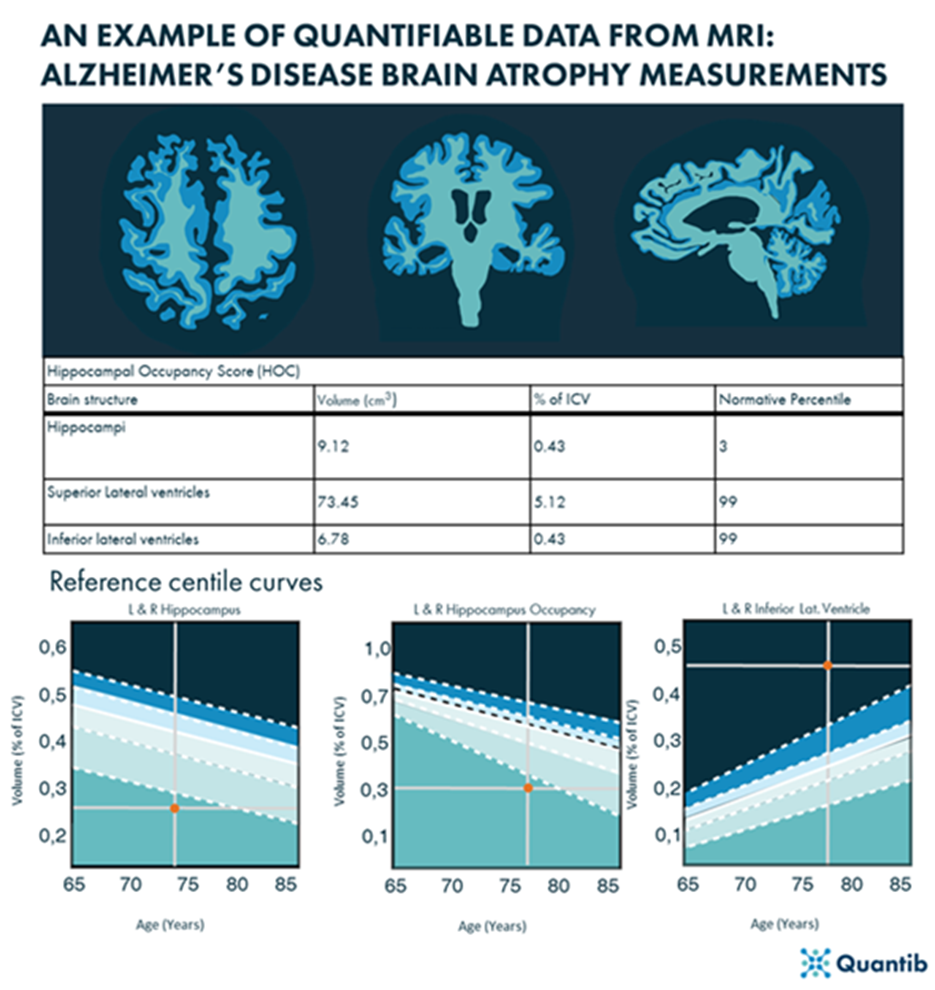
One of the most powerful tools for visualizing dementia is magnetic resonance imaging (MRI). MRI uses a strong magnetic field and radio waves to produce detailed images of the brain. This technology has revolutionized our understanding of dementia by allowing us to see the physical changes that occur in the brain as the disease progresses. For example, in the early stages of Alzheimer’s disease, MRI can reveal shrinkage of the hippocampus, a key region of the brain involved in memory and learning. As the disease advances, the brain’s overall volume may decrease, and other regions may become damaged.
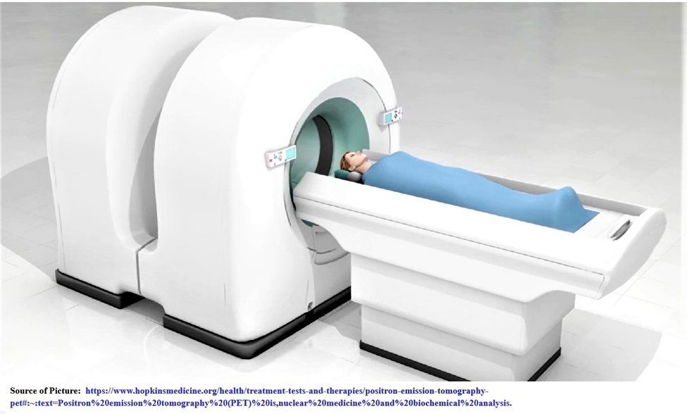
In addition to MRI, there are other imaging techniques that can be used to “visualize” dementia. Functional MRI can measure changes in blood flow in the brain and can be used to study how different brain regions are activated during certain tasks. This technology has helped researchers to understand how dementia affects specific cognitive functions, such as language, attention, and memory. Positron emission tomography (PET), a combination of nuclear medicine and biochemical analysis, is another imaging tool that can be used to diagnose dementia by measuring glucose and oxygen metabolism in the brain.
“PET works by using a scanning device (a machine with a large hole at its center) to detect photons (subatomic particles) emitted by a radionuclide in the organ or tissue being examined. PET differs from other nuclear medicine examinations in that PET detects metabolism within body tissues, whereas other types of nuclear medicine examinations detect the amount of a radioactive substance collected in body tissue in a certain location to examine the tissue’s function. Specifically, PET studies evaluate the metabolism of a particular organ or tissue, so that information about the physiology (functionality) and anatomy (structure) of the organ or tissue is evaluated, as well as its biochemical properties.”3/
Visualizing dementia can also involve the use of immersive technologies such as virtual reality. By creating 3D simulations of the brain, researchers can visualize and explore the complexities of dementia in a way that was previously impossible. For example, a virtual reality experience called “The Enemy Within” allows viewers to see what it’s like to experience dementia, from the inside out. This kind of visualization tool can help increase understanding and empathy among caregivers, family members, and the wider public.
Conclusion
Visualizing dementia through different imaging technologies has opened new avenues of research and treatment. The ability to see inside the brain and to identify specific changes that occur in the early stages of the disease has the potential to lead to earlier, more accurate diagnoses and more effective treatments. Developments in PET/CT and MRI hold great potential for visualizing and identifying dementia-specific signatures, possibly even before the onset of cognitive decline. While the disease itself may be invisible, our ability to “see” it is growing, and with it, our understanding and ability to respond to it. END
NOTES
1/ Tiago Beck, Visualizing Dementia: How Developments in CT and MRI Imaging help differentiate dementia subtype citing Prince, M. et al. The global prevalence of dementia: a systematic review and metaanalysis. Alzheimer’s & dementia 9, 63–75 (2013). https://www.quantib.com/blog/visualizing-dementia
2/ Tiago Beck, Visualizing Dementia: How Developments in CT and MRI Imaging help differentiate dementia subtype citing Steele, J. D. & Lawrie, S. M. 4 – Neuroimaging. in (eds. Johnstone, E. C., Owens, D. C., Lawrie, S. M., McIntosh, A. M. & Sharpe, M. B. T.-C. to P. S. (Eighth E.) 77–94 (Churchill Livingstone, 2010). doi:https://doi.org/10.1016/B978-0-7020-3137-3.00004-8..
3/ Positron Emission Tomography (PET), Johns Hopkins Medicine. https://www.hopkinsmedicine.org/health/treatment-tests-and-therapies/positron-emission-tomography-pet#:~:text=Positron%20emission%20tomography%20(PET)%20is,nuclear%20medicine%20and%20biochemical%20analysis.